10 things you need to know about the university's new micro-CT scanner
A micro-CT scanner is a fantastic research tool, says Associate Professor Henrik Lauridsen from the Department of Clinical Medicine. Learn more about this large machine, of which there are only a few in Denmark.

Let's celebrate!
Join us in celebrating the university's new scanner – and learn more about what it can do.
Come to the reception on Thursday, September 12th, from 2-4 PM.
Auditorium C, 114-101, Aarhus University Hospital.
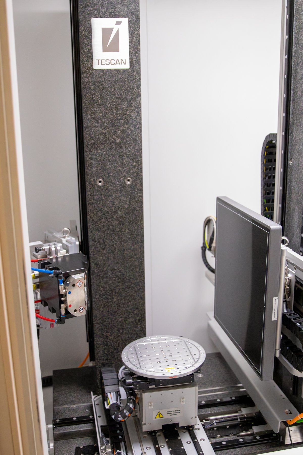
6.5 tons…
... is the weight of the scanner, which is encased in lead. This heavy casing ensures that it is completely safe to work in the room, as the lead shield protects against radiation hazards.
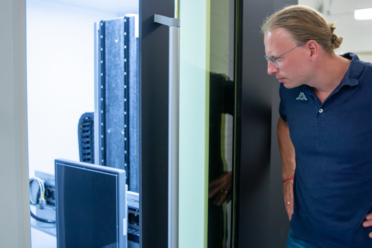
A micro-CT scanner…
... is quite large despite its name, at least the model located in Skejby. Several door frames had to be removed and reinstalled to move it into the basement of Aarhus University Hospital. The scanner uses X-rays to look inside an object, slice by slice. This can take a long time, but the 3D imaging technique provides extremely high resolution and great detail.
It is one of the few micro-CT scanners in Denmark…
... that can scan large objects, such as a femur from a large animal, at very high resolution. The scanner can accommodate samples up to 60 cm in diameter and up to 1 meter in height.
This makes it sought after by Danish museums and foreign researchers. Originally, the scanner was developed for mining purposes, such as analyzing whether there is gold in a soil sample.
The fetus of a minke whale…
... fits perfectly in the scanner. This became evident when Museum Sønderjylland wanted to scan a small minke whale from Greenland preserved in alcohol.
Other samples that have been scanned include forensic tissue samples, organs or bones from experimental animals, fossils like an ancient turtle, ceramics from the early Neolithic period, the head of the Tollund Man, soil samples (to reveal air-filled cavities, roots, biochar, compact zones, and other factors crucial for soil quality), and fossilized dinosaur droppings (to identify what the dinosaur ate).
In more serious cases, the scanner can also accommodate a deceased infant if the Department of Forensic Medicine wishes to develop new scanning methods to detect injuries suspected to be caused by violence.
315…
... scans have been performed since the micro-CT scanner arrived in November 2023 in the basement of the Department of Forensic Medicine, entrance D, at Aarhus University Hospital.
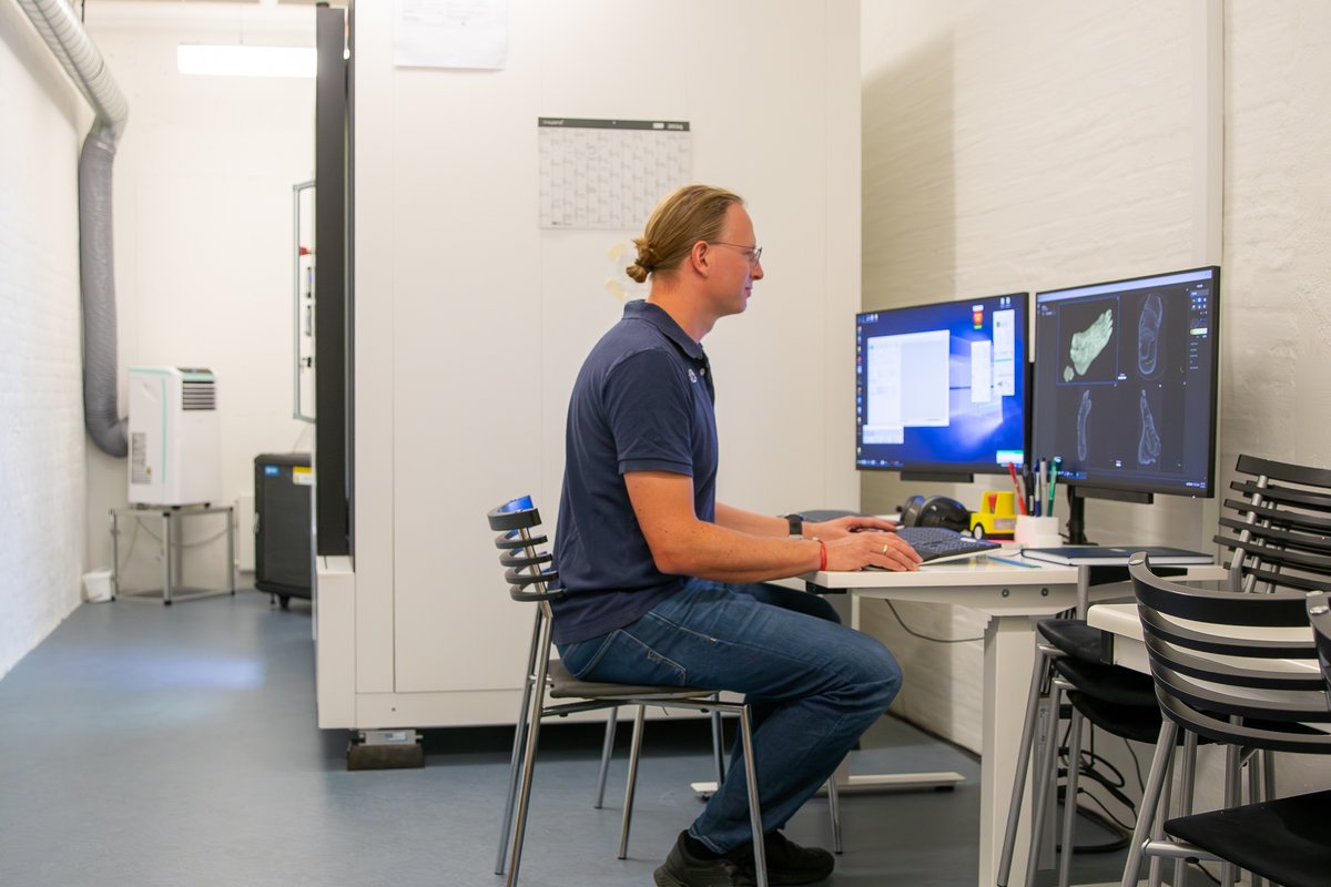
If a scan is to result in images with very high resolution, the process can take up to eight hours. Therefore, researchers often start the scanner before leaving for the day so that it can run overnight. However, it is also possible to conduct a full 360-degree dynamic scan in 10 seconds. This is useful for capturing a process that occurs relatively quickly, such as the development of gas bubbles in a chemical reaction or visualizing microanatomical changes in a bone under stress.
The X-ray source…
... can range from 30 kV to 180 kV. This means the scanner can penetrate hard materials like bones, teeth, metallic implants, soil samples, stones, and fossils, and it can produce highly detailed images of soft tissues like organs. A typical CT scanner has an X-ray voltage range of 80 to 140 kV, but because the energy level of the micro-CT scanner can be precisely adjusted to the scanned object, there is often improved contrast between different structures in the object.
The scanner is not ideal for…
... amber containing dinosaur feathers or insect skeletons because everything in such a sample is carbon-based, resulting in a gray-on-gray image. The scanner works best when there are different materials that provide good contrast. However, for soft tissues, such as a euthanized experimental animal, contrast can still be achieved using different contrast agents that penetrate and create X-ray density differences between various structures.
A hair is about 100 µm thick…
... and the scanner can produce images with a resolution down to 2 µm. This means it can easily show detail at the muscle fiber level and reveal how well a small rat bone has healed after a fracture.
The Czech/Belgian company TESCAN
... manufactured the large machine. The scanner was purchased through an interdisciplinary collaboration between the Department of Clinical Medicine, the Department of Forensic Medicine, and the Department of Agroecology. Most of the funding was provided by the Carlsberg Foundation and the Department of Agroecology.
Henrik Lauridsen and the other researchers involved with the scanner now dream of upgrading it to enable hyperspectral imaging, a technique that uses the electromagnetic spectrum to identify materials in an object down to the elemental level. This would allow the scanner to determine, for example, if a sample contains lead fragments (for forensic gunshot investigations) and add "color" to otherwise gray images, as seen with histochemical staining by infiltrating soft tissue or organic matter in soil with various dissolved metal salts.
Just reach out…
... to Henrik, Kasper, or Lars if you think the scanner could elevate your research to new heights. See their contact details below. For now, it costs nothing to try the machine, but it may eventually be necessary to introduce a fee to cover service costs.
Contact:
Associate Professor Henrik Lauridsen
Aarhus University, Department of Clinical Medicine
Phone: +45 61 72 21 06
Email: henrik@clin.au.dk
Assistant Professor Kasper Hansen
Aarhus University, Department of Forensic Medicine
Phone: +45 29 70 25 80
Email: kaha@forens.au.dk
Professor Lars Juhl Munkholm
Aarhus University, Department of Agroecology - Soil Biology and Nutrients
Phone: +45 25 15 27 16
Email: lars.munkholm@agro.au.dk